Human brain anatomy explains how the brain is built, how its different parts are arranged, and how those parts work together. The brain is one of the most remarkable organs in the human body. It allows us to think, learn, remember, speak, move, feel emotions, understand our surroundings, and control many activities that happen without conscious effort.
Although the brain may look like a single soft organ, it contains many specialized regions. Each region has important responsibilities, but no part works completely alone. Brain cells constantly communicate with one another through complex networks.
Learning about human brain anatomy doesn’t have to be difficult. Once the main structures are understood, the subject becomes much easier to follow. The cerebrum, cerebellum, brainstem, cerebral cortex, brain lobes, thalamus, hypothalamus, hippocampus, and other structures all have different roles.
This guide explains these structures in simple language and shows how they work together to support everyday human life.
What Is the Human Brain?
The brain is the main control center of the nervous system. Along with the spinal cord, it forms the central nervous system. It receives information from the senses, processes that information, and helps the body respond.
The brain is protected by the skull, membranes called meninges, and cerebrospinal fluid. It also receives a continuous supply of oxygen and nutrients through the blood.
The adult human brain weighs roughly 1.3 to 1.4 kilograms on average, although brain size varies naturally between individuals. More important than its size is its organization. Billions of nerve cells and supporting cells communicate through an enormous network.
A useful way to understand human brain anatomy is to imagine the brain as a busy communication center. Different departments have different jobs, but they constantly share information.
For example, when you pick up a glass of water, your brain must recognize the glass, understand its position, plan the movement, control your muscles, and adjust your hand as it approaches the object. Several brain regions participate in this simple action.
The Main Parts of the Brain
The brain is commonly divided into three large areas:
- The cerebrum
- The cerebellum
- The brainstem
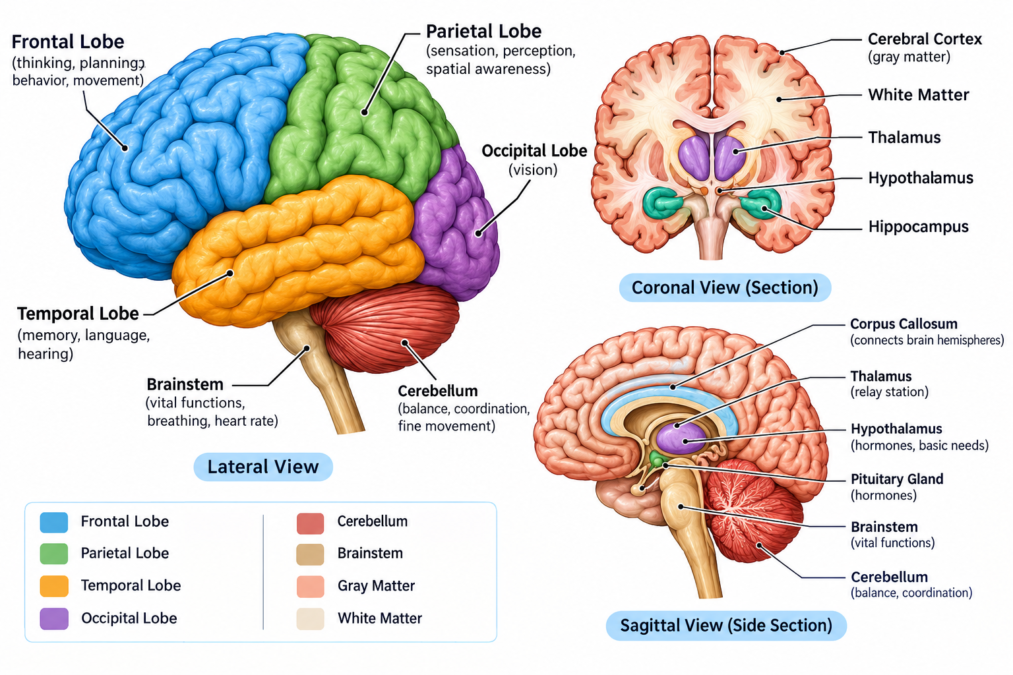
The cerebrum is the largest part of the brain. It is involved in thinking, memory, language, sensation, and voluntary movement.
The cerebellum is located toward the back and lower part of the brain. It helps coordinate movement, balance, posture, and motor learning.
The brainstem connects the brain with the spinal cord. It is involved in important functions such as breathing, heart rate, blood pressure, swallowing, and alertness.
However, this simple division doesn’t tell the whole story. Deeper structures such as the thalamus, hypothalamus, hippocampus, amygdala, and basal ganglia also play important roles.
Understanding these major divisions gives us a strong foundation for studying human brain anatomy.
| Brain Structure | Main Location | Major Functions |
|---|---|---|
| Cerebrum | Upper and largest part | Thinking, memory, language, sensation, movement |
| Cerebellum | Back and lower brain | Balance, coordination, motor learning |
| Brainstem | Lower brain | Breathing, heart rate, alertness, communication with spinal cord |
| Thalamus | Deep central region | Sensory and motor information processing |
| Hypothalamus | Below the thalamus | Temperature, hunger, thirst, hormones |
| Hippocampus | Temporal region | Learning and memory |
| Amygdala | Deep temporal region | Emotional processing |
| Basal ganglia | Deep cerebral regions | Movement and other brain functions |
The Cerebrum and Its Two Hemispheres
The cerebrum makes up most of the brain. It has two large halves called the left hemisphere and right hemisphere.
The two hemispheres are separated by a deep groove called the longitudinal fissure. They communicate with each other through a large bundle of nerve fibers known as the corpus callosum.
The hemispheres have some functional differences. For example, language functions are often more strongly represented in the left hemisphere in people who are left-language dominant. However, it would be misleading to say that one side performs all logical tasks while the other handles all creative tasks.
In reality, both sides communicate constantly.
The hemispheres also generally control movement on opposite sides of the body. The left side of the brain controls many movements on the right side, while the right side controls many movements on the left side.
The outer surface of the cerebrum is called the cerebral cortex. It contains gray matter and is divided into different regions.
The surface contains folds called gyri and grooves called sulci. These folds allow a large amount of cortical tissue to fit inside the skull.
This folded structure is one of the most recognizable features of human brain anatomy.
The Four Main Lobes of the Brain
The cerebral hemispheres are traditionally divided into four major lobes:
- Frontal lobe
- Parietal lobe
- Temporal lobe
- Occipital lobe
These lobes have different areas of specialization, although their functions overlap. Complex activities usually depend on several regions working together.
The Frontal Lobe
The frontal lobe is located behind the forehead. It is involved in planning, decision-making, problem-solving, attention, voluntary movement, and several aspects of behavior.
It also contains the primary motor cortex, which helps control voluntary movements.
In the dominant hemisphere, parts of the frontal lobe are involved in speech production. The frontal lobe also contributes to higher-level thinking and the ability to plan actions.
For example, if you’re preparing a meal, your brain may need to remember the recipe, decide what to do first, control your hands, and monitor the result. The frontal lobe participates in many of these processes.
The Parietal Lobe
The parietal lobe is located behind the frontal lobe. It is strongly involved in processing sensory information from the body.
It helps the brain understand sensations such as:
- Touch
- Pressure
- Temperature
- Pain
- Body position
The parietal lobe also contributes to spatial awareness. It helps us understand where our body is in relation to objects around us.
If you reach for a phone on a table, your brain needs to understand both the position of your hand and the position of the phone. Several systems, including the parietal regions, help make this possible.
The Temporal Lobe
The temporal lobe is located on the sides of the brain. It plays an important role in hearing, language understanding, memory, and emotional processing.
Auditory information is processed in areas of the temporal lobe. Other parts are involved in understanding spoken language and recognizing meaningful sounds.
Deep inside the temporal region are structures such as the hippocampus and amygdala.
Because the temporal lobe is involved in so many important processes, it is an important part of human brain anatomy.
The Occipital Lobe
The occipital lobe is located at the back of the brain and is strongly associated with vision.
The eyes collect visual information, but the eyes don’t actually “understand” what they see. Instead, signals travel through visual pathways to the brain, where information about shapes, colors, movement, and other visual features is processed.
The occipital lobe therefore plays a central role in turning visual signals into meaningful information.
The Cerebral Cortex
The cerebral cortex is the outer layer of the cerebrum. It contains billions of neurons and supports many complex functions.
Different areas of the cortex are associated with different types of processing. Some areas receive sensory information, while others control movement or participate in language, memory, attention, and reasoning.
The cortex can be divided into functional areas, including:
- Motor areas
- Sensory areas
- Visual areas
- Auditory areas
- Language-related areas
- Association areas
Association areas are especially important because they combine information from different systems.
For instance, recognizing a familiar person isn’t simply about seeing a face. The brain may connect visual information with memories, emotions, names, and previous experiences.
This cooperation is a central idea when studying human brain anatomy.
The Cerebellum and Coordination
The cerebellum is located at the back of the brain, underneath the occipital and temporal regions of the cerebrum.
It is strongly involved in coordination, balance, posture, and motor learning. It helps make movements smooth and accurate.
Think about learning to ride a bicycle. At first, maintaining balance may require considerable attention. With practice, movements become more natural. The cerebellum is part of the system that helps the brain learn and refine such motor skills.
The cerebellum also receives information from the body and other parts of the nervous system. It compares information and helps adjust movements.
Although it has traditionally been associated mainly with movement, research also suggests that the cerebellum participates in some cognitive processes.
The Brainstem and Its Essential Functions
The brainstem connects the brain to the spinal cord. It is located beneath the cerebrum and in front of the cerebellum.
The three main parts of the brainstem are:
- Midbrain
- Pons
- Medulla oblongata
The midbrain is involved in movement, sensory processing, eye movements, and certain reflexes.
The pons connects different parts of the nervous system and has roles in movement, sensation, sleep, breathing, and facial functions.
The medulla oblongata is the lowest part of the brainstem. It contains important centers involved in breathing, heart activity, blood pressure, swallowing, and other automatic functions.
Because of these responsibilities, the brainstem is a critical part of human brain anatomy.
The Thalamus and Hypothalamus
Some important brain structures are located deep beneath the cerebral cortex.
The thalamus acts as an important relay and processing center. Much of the sensory information traveling toward the cerebral cortex passes through the thalamus. It also participates in motor and other brain networks.
The hypothalamus is smaller but has an extremely important role in maintaining the body’s internal balance.
It helps regulate:
- Body temperature
- Hunger
- Thirst
- Sleep-related processes
- Hormone regulation
- Stress responses
- Other automatic body functions
The hypothalamus works closely with the pituitary gland and endocrine system.
Although these structures are not visible when looking at the outer surface of the brain, they are essential when studying human brain anatomy.
The Hippocampus and Memory
The hippocampus is a structure located deep within the temporal region of the brain.
It plays an important role in learning and memory. It is particularly important for forming and organizing certain types of new memories.
Suppose you visit a new city for the first time. You may remember the streets, buildings, sounds, and experiences from that visit. Several brain systems contribute to this memory, including the hippocampus.
The hippocampus doesn’t work alone. Memory involves networks distributed throughout the brain.
This is important because popular explanations sometimes make it seem as if one small structure is responsible for all memory. The real situation is much more complex.
The Amygdala and Emotional Processing
The amygdala is a small group of structures located deep within the temporal region.
It plays an important role in processing emotionally significant information. It is especially well known for its involvement in fear and threat-related responses, although its role is broader than fear alone.
Emotions can influence attention, memory, learning, and behavior. That’s why an emotional event may remain especially memorable.
The amygdala works with other brain regions rather than operating independently. Its connections with the hypothalamus, hippocampus, and cerebral cortex help link emotions with memories and physical responses.
Gray Matter and White Matter
Two terms that frequently appear in discussions of human brain anatomy are gray matter and white matter.
Gray matter contains many neuron cell bodies and is found in the cerebral cortex and several deeper brain structures.
White matter contains many nerve fibers, especially axons that connect different parts of the nervous system. Many of these axons are covered by myelin, a fatty substance that helps signals travel efficiently.
A simple comparison is useful:
Gray matter:
Often involved in processing information.
White matter:
Provides many communication pathways between different brain areas.
This doesn’t mean gray matter and white matter have completely separate jobs. They depend on each other.
A processing center is only useful if it can communicate with other centers. Likewise, communication pathways are useful because they connect areas that perform important functions.
The Ventricles and Cerebrospinal Fluid
The brain contains four connected spaces called ventricles. These spaces are filled with cerebrospinal fluid, commonly known as CSF.
The ventricular system includes:
- Two lateral ventricles
- The third ventricle
- The fourth ventricle
CSF also circulates around the brain and spinal cord.
It helps protect the nervous system and provides a controlled environment around the brain and spinal cord. It also participates in the movement of certain substances through the central nervous system.
The ventricular system is another important feature of human brain anatomy because it shows that the brain contains an organized internal fluid system, not simply solid tissue.
How the Brain Is Protected
The brain is extremely delicate, so the body provides several layers of protection.
The skull is the first major protective structure. Under the skull are three membranes known as the meninges.
They are:
- Dura mater
- Arachnoid mater
- Pia mater
The dura mater is the strong outer layer. The arachnoid mater lies underneath it, while the pia mater closely follows the surface of the brain.
Cerebrospinal fluid also provides cushioning around the brain and spinal cord.
Together, the skull, meninges, and cerebrospinal fluid help protect the nervous system from physical stress.
Blood Supply to the Brain
The brain requires a constant supply of oxygen and glucose. It receives blood through an extensive network of arteries.
Important arteries include the:
- Anterior cerebral arteries
- Middle cerebral arteries
- Posterior cerebral arteries
Other blood vessels also contribute to the brain’s circulation.
The brain has a high energy demand. Even though it makes up only a small percentage of total body weight, it uses a significant amount of the body’s energy.
If blood flow to part of the brain is interrupted, brain tissue can become damaged. This is one reason strokes require urgent medical treatment.
Blood supply is therefore a key topic in human brain anatomy and clinical medicine.
Neurons: The Communication Cells of the Brain
Neurons are specialized cells that communicate information throughout the nervous system.
A typical neuron includes:
- Cell body
- Dendrites
- Axon
Dendrites receive information, while the axon carries signals away from the cell body.
Neurons communicate with other cells through structures called synapses. Chemical messengers called neurotransmitters can carry information across many synapses.
The brain also contains glial cells. These cells support neurons in several ways, including maintaining the environment around them and helping with protection and repair.
The enormous communication network formed by neurons and supporting cells makes the brain capable of processing a huge amount of information.
How Different Brain Areas Work Together
One of the biggest lessons from human brain anatomy is that complex activities usually depend on networks rather than a single brain structure.
Consider reading a simple sentence.
First, the eyes receive light and send signals through the visual system. Visual areas process the shapes of letters. Language-related areas help identify words and meaning. Memory systems connect the information with knowledge you already have.
Now consider walking.
Your brain needs information about balance, body position, the environment, muscle movement, and the direction you want to travel. Different brain systems communicate continuously to keep your movements controlled.
Even something as simple as picking up a cup involves vision, attention, movement planning, muscle control, and sensory feedback.
The brain is constantly coordinating these processes, often without conscious awareness.
Human Brain Anatomy in Everyday Life
You don’t need to be a neuroscientist to see how the brain works. Everyday activities provide countless examples.
When you listen to music, auditory areas process sound while memory and emotional networks may respond to familiar melodies.
When you learn a new language, the brain creates and strengthens connections involved in hearing, speaking, reading, memory, and meaning.
When you play a sport, the brain coordinates vision, movement, balance, timing, and decision-making.
When you study for an exam, attention, memory, language, and reasoning systems work together.
When you meet someone you know, your brain processes the person’s face, voice, name, memories, and emotional associations.
These examples make human brain anatomy easier to understand because they connect anatomical structures with real experiences.
Why Understanding Brain Anatomy Matters
Learning about the brain is important for both education and medicine.
Doctors and scientists use knowledge of brain structure to understand neurological conditions, injuries, and diseases.
Brain anatomy is especially relevant when studying conditions such as:
- Stroke
- Epilepsy
- Brain tumors
- Traumatic brain injury
- Movement disorders
- Memory disorders
- Neurological infections
Medical imaging, including MRI and other techniques, allows healthcare professionals to examine brain structures and identify abnormalities.
However, brain images must be interpreted by trained professionals. A scan is only one part of a complete medical assessment.
Common Myths About the Human Brain
The brain is surrounded by many popular myths. One famous claim says that people use only a small percentage of their brains. This isn’t an accurate description of how the brain works.
Another common myth suggests that people are completely “left-brained” or “right-brained.” Although the two hemispheres have some functional differences, they work together through extensive communication pathways.
It’s also misleading to think that every mental ability exists in one small location. Some functions are strongly linked with particular areas, but complex activities often involve networks across different regions.
These facts remind us that human brain anatomy is much more complicated than simple diagrams or social-media claims might suggest.
A Simple Table of Major Brain Functions
The following table provides an easy way to review the major structures:
| Part | Main Role |
|---|---|
| Frontal lobe | Planning, movement, attention, decision-making |
| Parietal lobe | Body sensations and spatial processing |
| Temporal lobe | Hearing, language, memory |
| Occipital lobe | Visual processing |
| Cerebellum | Coordination, balance, motor learning |
| Brainstem | Vital automatic functions and communication with spinal cord |
| Thalamus | Sensory and motor information processing |
| Hypothalamus | Internal body regulation |
| Hippocampus | Learning and memory |
| Amygdala | Emotional processing |
| Cerebral cortex | Higher-level processing and conscious functions |
| Corpus callosum | Communication between cerebral hemispheres |
This table doesn’t cover every structure in the brain, but it provides a useful starting point for anyone beginning to study human brain anatomy.
Final Thoughts on Human Brain Anatomy
The human brain is an extraordinary organ made up of many connected structures. The cerebrum supports thinking, memory, language, sensation, and voluntary movement. The cerebellum helps coordinate movement and balance, while the brainstem controls or supports many functions that are essential for life.
Deeper structures such as the thalamus, hypothalamus, hippocampus, and amygdala add further layers of control and processing. Meanwhile, neurons and glial cells create the communication system that allows all these regions to work together.
Perhaps the most interesting part is that the brain isn’t simply a collection of separate pieces. Its real power comes from communication between regions. Every conversation, movement, memory, emotion, and new skill depends on networks working together.
As neuroscience continues to develop, researchers are learning more about how the brain changes with age, learning, experience, injury, and disease. There is still much to discover.
For anyone interested in medicine, psychology, education, or simply understanding how the human body works, human brain anatomy provides an excellent foundation. The more we learn about this complex organ, the better we can appreciate how remarkable the human body really is.
Stay in touch to get more updates & news on See Blog Hub!